Close
Select your language

The page doesn't exist in this language

Find your Coloplast country website

Introduction

Get a useful overview of the key issues in ostomy surgery in this section. There is a quick reference to the anatomy and organ systems involved, the common complications that your patients may encounter after surgery and tips and tools to help. 


Set your patients up for success

Ostomate shopping for groceries

Assessing quality of life in ostomy patients

The Stoma Quality of Life tool (Stoma-QoL) is a simple, validated questionnaire that can be used to measure quality of life in people with a stoma – and identify the issues that may cause anxiety or concern. Stoma Quality of Life tool
Close

Assessing quality of life in ostomy patients

People with an ostomy can have many concerns – fear of leakage, embarrassment about their body, worries about becoming a burden to family and friends. When these concerns stop people from doing the things they enjoy, it puts their quality of life at risk.

With the Stoma-QoL tool, healthcare professionals now have a standard and a common language with which to assess quality of life for people with a stoma. You can use the tool to monitor quality of life over time in the same person – or compare quality of life between patients.

Stoma-QoL is:

  • Specifically designed for people with a stoma – all questions are based on input from people with a stoma
  • Validated – tested in representative ostomy populations in different countries
  • Reliable – weighted to emphasize the issues that are most critical to the respondent’s quality of life
  • Cross-cultural – translated into 16 languages
  • Simple – the questionnaire only takes 5–10 minutes to complete

Download the tool:

English

French

Close
Surgical education pad

Surgical education pad

Printable note pages can help explain specific surgical procedures and anatomy to pre-op patients. Download the surgical education pad
Close

Helpful tools to support your work

Surgical Education Pad
Understanding the basics of a surgical procedure is important for many patients, as it can help them feel prepared and better able to cope. The Surgical Education Pad is an educational tool for proactive patients who would like detailed information about their surgery.

The file includes information on:

  • Abdomino-perineal excision of rectum (APER)
  • Low anterior resection
  • Hartmann’s procedure
  • Total colectomy
  • Restorative proctocolectomy
  • Pan-proctocolectomy
  • IIeal conduit
Close

Basic anatomy

The digestive system

The digestive system

The digestive system is one of the body's major organ systems. The digestive tract handles the digestion and processing of food. More about the digestive system
Close

Anatomy and physiology of the digestive and urinary systems

Digestive system

 
The digestive tract stretches some nine metres from the mouth to the anus and is divided into different sections. Each section processes food in a specific way to prepare it for the next section of the digestive tract, until the waste finally leaves the intestinal tract as faeces.

Functions of the digestive system:

  • Mechanical and chemical breakdown of food into basic nutrients
  • Absorption of nutrients into the blood
  • Processing and elimination of waste

A number of organs work alongside the digestive tract, producing fluids and enzymes to aid indigestion:

  • Salivary glands in the mouth
  • Acid fluids in the stomach
  • Liver and gallbladder
  • Pancreas


The gastrointestinal tract comprises:

  • Mouth
  • Oesophagus
  • Stomach
  • Small intestine – jejunum & ileum
  • Large intestine – colon
  • Rectum
  • Anus


The intestinal wall consists of several layers


The small intestine is approximately 5–7 m long in adults. It is divided into three main parts:

  • Duodenum
  • Jejunum
  • Ileum

The jejunum and ileum are connected to the abdominal wall by the mesentery. The mesentery contains arteries, veins and lymph vessels that ensure the transport of oxygen and nutrients to and from the small intestine.


The large intestine is approximately 1–2 m long in adults. It is divided into six parts:

  • Ascending colon
  • Transverse colon
  • Descending colon
  • Sigmoid colon, the S-shaped structure
  • Rectum: final part of the digestive tract. Stool collects in the rectal ampulla. A filled ampulla initiates the urge to empty the bowels
  • Anus, terminal opening of the digestive system
Close
The urinary system

The urinary system

The urinary system is another of the body's major organ systems. The urinary tract is involved in fluid and electrolyte balance, and the excretion of urine. More about the urinary system
Close

The urinary system

The urinary system

The overall function of the urinary system is to produce and drain urine, removing waste products and regulating the blood's fluid balance. The entire urinary system is located behind the digestive tract.


The urinary system consists of:

  • Two kidneys
  • Two ureters
  • Bladder
  • Urethra

The kidneys are two bean-shaped structures that continuously filter the blood, removing waste products and excess water, and balancing fluids and electrolytes. This filtering process results in the production of urine.


The ureters are ducts that carry urine from the kidneys to the bladder.


The bladder has a dual function. It is both a reservoir that stores urine and a pump that expels urine from the body. The muscle in the bladder wall pushes the urine out.


The urethra is a duct connecting the bladder to the outside of the body.

Close

Basic surgical procedures

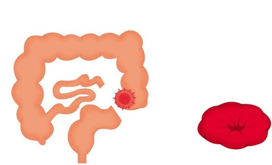
Location of a colostomy

Colostomy surgery

A colostomy is a surgically created opening in part of the large intestine which can be permanent or temporary, depending on the disease process. Colostomy surgery
Close

Colostomy surgery

Formation of a stoma

Colostomy

In a colostomy operation, part of the colon is brought to the surface of the abdomen to form the stoma. A colostomy is usually (but not always) created on the left-hand side of the abdomen. Output consistency depends on where the colostomy is located:

  • Ascending colostomy: Output can range from liquid to pasty consistency and may be irritating to the skin
  • Transverse colostomy: Output is somewhat formed
  • Descending / sigmoid colostomy: Output is formed


Because a stoma has no muscle to control defecation, the output it produces will need to be collected using a stoma pouch.


There are two different types of colostomy surgery: End colostomy and loop colostomy.


End colostomy
If parts of the large intestine (colon) or rectum have been removed, the remaining large intestine is brought to the surface of the abdomen to form a stoma. An end colostomy can be temporary or permanent. The temporary solution is relevant in situations where the diseased part of the bowel has been removed and the remaining part of the bowel needs to rest before the ends are joined together. The permanent solution is chosen in situations where it is too risky or not possible to reconnect the two parts of the intestine.


Loop colostomy
In a loop colostomy, the bowel is lifted above skin level and held in place with a stoma rod. A cut is made on the exposed bowel loop, and the ends are then rolled down and sewn onto the skin. In this way, a loop stoma actually consists of two stomas (double-barrelled stoma) that are joined together. The loop colostomy is typically a temporary measure performed in acute situations. It can also be carried out to protect a surgical join in the bowel.

Close
Location of a ileostomy

Ileostomy surgery

An ileostomy is a surgically created opening in the small intestine called the ileum with liquid to pasty consistency. Ileostomy surgery
Close

Ileostomy surgery

Ileostomy

In an ileostomy operation, a part of the small intestine called the ileum is brought to the surface of the abdomen to form the stoma. Ileostomy surgery is typically performed in cases where the end part of the small intestine is diseased, and is usually made on the right-hand side of the abdomen. Depending on the disease process, ileostomies may be permanent or temporary.


The stool can range from a liquid to pasty consistency, and contains enzymes that are irritating to the peristomal skin. Because a stoma has no muscle to control defecation, the output will need to be collected in a pouch.


There are two different types of ileostomy surgery:

End ileostomy

End ileostomy

An end ileostomy is created when part of the large intestine (colon) is removed (or simply needs to rest) and the end of the small intestine is brought to the surface of the abdomen to form a stoma. An end ileostomy can be temporary or permanent.

The temporary solution is relevant in situations where the diseased part of the bowel has been removed and the remaining part needs to rest before the ends are joined together. The permanent solution is chosen in situations where it is too risky or not possible to reconnect the two parts of the intestine.

Loop ileostomy

Loop ileostomy

In a loop ileostomy, a loop of the small intestine is lifted above skin level and held in place with a stoma rod. A cut is made on the exposed bowel loop, and the ends are then rolled down and sewn onto the skin. In this way, a loop ileostomy actually consists of two stomas that are joined together.

The loop ileostomy is typically temporary and performed to protect a surgical join in the bowel. If temporary, it will be closed or reversed in a later operation.


Close
Location of a urostomy

Urostomy surgery

A urostomy / ileal conduit is created from a piece of small intestine. The ureters are surgically tunneled into a small segment of the small intestine called a "conduit" or "pipeline". Urostomy surgery
Close

Urostomy surgery

UrostomyUrostomy

If the bladder or urinary system is damaged or diseased and your patient is unable to pass urine normally, there is a need for a urinary diversion. This is called a urostomy, an ileal conduit or a Bricker bladder.

An isolated part of the intestine is brought onto the surface of the right-hand side of the abdomen and the other end is sewn up. The ureters are detached from the bladder and reattached to the isolated section of the intestine. Because this section of the intestine is too small to function as a reservoir, and there is no muscle or valve to control urination, your patient will need a urostomy pouch to collect the urine.

Close

Complications with a stoma

Peristomal skin disorders

Peristomal skin issues are the most common complications for people with a stoma, accounting for a third of visits to stoma care nurses. Most frequent cause is chemical irritation due to leakage. Tools to help accurate assessment and appropriate treatment
Close

Peristomal skin disorders

Monitoring of peristomal skin, already during the post-operative phase, is essential to ensure that the skin remains healthy, and, if it is affected by a skin disorder, that appropriate and consistent treatment can be initiated immediately.


The Ostomy Skin Tool is developed together with stoma care nurses on the Global Advisory Board to ensure accurate assessment of peristomal skin.


It consists of two parts; the DET score and the AIM guide:

  • DET score (Discolouration, Erosion, Tissue overgrowth) – provides a standardised and validated way to score the peristomal skin through objective observations.
  • AIM guide (Assessment, Intervention, Monitoring)  - provides categorisation of the peristomal skin disorder according to its cause and offers guidance on care. The guide is based on a thorough literature review.


The tool is designed to help you to:

  • Assess peristomal skin at the time of assessment based on the validated DET score
  • Identify the most suitable appliance and peristomal skin care routine
  • Provide you and your colleagues with a common language for describing peristomal skin conditions 
Close

61% of patients experience peristomal skin conditions

We learned from the DialogueStudy that 61% of patients have a peristomal skin condition. Here's an overview of prevention and management for some of the most common peristomal skin conditions. Common peristomal skin conditions
Close

Peristomal Skin Conditions

HyperplasiaHyperplasia (Pseudoverrucous Lesions)

Most often seen with urostomies. The wart-like lesions are usually caused by urine pooling on the skin for extended periods of time.


Prevention:

  • Correctly cut the barrier to the size of the stoma
  • Assess for leakage. Examine ostomy barrier for erosion upon removal and adjust wear time accordingly
  • Consider using an extended wear barrier
  • If stoma is flush or retracted, consider a pouching system with convexity
  • Use a pouch that has a built-in anti-reflux valve to prevent urine from washing over the stoma
  • Use a bedside drainage system at night

Management:

  • Consult physician or ET Nurse
  • Identify the underlying cause
  • Modify the pouching system
  • Check urine pH to assess urine acidity
  • Physician’s orders may include white vinegar soaks and/or a Colly-Seel-type barrier to the wart-like lesions until the condition improves


CandidiasisFungal Infection (Candida / Yeast)

Candida is a common skin flora that grows in dark, damp sites such as under an ostomy barrier. The rash starts out as pustules before turning into a raised area with erythema consisting of irregular margins with surrounding satellite lesions. Patients may complain of itching or burning. Predisposing factors include antibiotic therapy, diabetes or immunosuppression.


Prevention:

  • Use a properly fitting pouching system 
  • Eliminate cause of moisture: inspect pouching system for signs of leakage
  • After bathing, dry the skin and the pouching system thoroughly
  • Assess wear time by examining the barrier for erosion upon removal

Management:

  • Consult with physician or ET Nurse
  • Identify the underlying cause
  • If skin is moist and weepy, consider the crusting technique using antifungal powder in place of ostomy powder (first obtain physician's order / prescription)
  • Systemic treatment may be prescribed by the physician if more than one body area is involved
  • Blood sugar management may also be considered


FolliculitisFolliculitis

Folliculitis is an inflammation of a hair follicle. It is often caused by bacterial sources such as staphylococcus aureus, streptococci and pseudomonas aeruginosa. Predisposing factors include antibiotic therapy, diabetes and immunosuppression.


Prevention:

  • Gently remove the barrier to prevent skin trauma
  • Shave hair in the direction of hair growth, but always away from the stoma
  • Use an electric razor
  • Wash, rinse and completely dry skin before applying a new pouching system

Management:

  • Consult with physician or ET Nurse
  • Identify the underlying cause
  • Avoid shaving hair in affected area - clip hair only
  • If skin is moist and weepy, consider the crusting technique using ostomy powder or an antifungal powder (first obtain physician’s order/prescription) if a fungal rash is present


Mechanical InjuryMechanical Injury

Mechanical injury is peristomal skin damage or skin stripping due to pressure, friction and/or shear. Contributing factors include: traumatic removal of the barrier, vigorously scrubbing the peristomal skin and/or a poorly fitting pouching system.


Prevention:

  • Gently clean the peristomal skin
  • Remove the barrier in the direction of hair growth using the push-pull technique
  • Use warm water or adhesive remover to remove the barrier if needed
  • Evaluate pouching system to ensure proper fit

Management:

  • Consult with physician or ET Nurse
  • If skin is moist and weepy, consider the crusting technique


Irritant Contact DermatitisIrritant Contact Dermatitis

Hypersensitivity to chemical agents such as stoma output, soaps and/or adhesives resulting in an inflammatory response. Associated with well-defined erythema, edema or loss of epidermis. Pruritus, crusting, oozing or dryness may be present. 


Prevention:

  • Measure and cut opening of barrier to the size of the stoma
  • Change barrier on schedule
  • Change barrier immediately if burning, irritation or signs of leakage occur
  • Examine the barrier for areas of erosion upon removal
  • Limit the use of products on the skin

Management:

  • Consult with physician or ET Nurse
  • Identify the underlying cause
  • Use correctly sized barrier and consider using convexity or belt
  • Consider using an extended wear barrier
  • If skin is moist and weepy, consider using crusting technique


Allergic Irritant DermatitisAllergic Irritant Dermatitis

Immunologic response due to exposure to an allergen. Associated with areas of erythema that may correspond to the shape of the contact surface.


Prevention:

  • Limit the use of products on the skin
  • Add one new product at a time to assess patient’s reaction

Management:

  • Consult with physician or ET Nurse
  • Remove known or suspected allergen - change type of pouching system and/or eliminate any unnecessary products
  • If skin is moist and weepy, consider crusting technique


Pyoderma GangrenosumPyoderma Gangrenosum

Rare inflammatory skin disorder with unknown etiology seen in patients with inflammatory bowel disease. The lesions are ulcerated with dusky red to purplish margins. Lesions can be painful.


Management:

  • Consult with physician or ET Nurse
  • Manage the underlying disease and infection per physician order
  • Manage ulcer pain per physician’s orders
  • Consider steroids with physician’s orders
  • Use a flexible barrier with a gentle adhesive
  • If skin is moist and weepy, consider crusting technique
  • Wound care may be ordered by the physician and is determined by amount of drainage and depth of wound
Close

Stomal challenges

In addition to the peristomal skin, stomal conditions can present challenges to a good seal, too. Common stomal challenges
Close

Common stomal challenges

ProlapseProlapse

A prolapse occurs when the bowel telescopes through the stoma, causing the stoma to increase in length. It is most common with loop colostomies. Contributing factors include: abdominal wall opening larger than the bowel, increased abdominal pressure and weak abdominal tone.


Management:

  • Consult with physician or ET Nurse
  • Instruct patient to avoid weight gain and suggest regular exercise to increase abdominal tone
  • Consider the use of prolapse support binder
  • Revise pouching system
  • Larger pouch to accommodate increased stoma length
  • Measure stoma base while stoma is protruding at its largest size (sitting position)
  • Cut barrier opening to accommodate stoma at its largest size
  • Use a flexible, flat barrier
  • Instruct patient to notify physician for signs and symptoms of obstruction and ischemia


Parastomal Hernia

Parastomal Hernia

Occurs due to a weakness in the muscle layer of the abdominal wall, allowing intestine to come through the muscle. Contributing factors include a too-large fascial opening around the stoma, poor muscle tone and placement of the stoma outside the rectus muscle.


Management:

  • Consult with physician or ET Nurse
  • Measure the stoma while patient is sitting up and stoma is at its largest
  • Use a one-piece pouching system or a two-piece adhesive coupling system, which allows flexibility and adapts to abdominal contours
  • Consider a hernia support belt for added support
  • Instruct patient to avoid excessive weight gain 


Mucocutaneous SeparationMucocutaneous Separation

The sutured junction between the stoma and the skin is called the mucocutaneous junction. When the junction completely or partially separates from the skin, it is called a mucocutaneous separation. Contributing factors include infection, tension on the suture line and delayed healing due to disease processes or corticosteroids.


Management:

  • Consult with physician or ET Nurse
  • Wound care may be ordered by the physician and is determined by amount of drainage and depth of wound
  • Change pouching system as needed to provide wound care


NecrosisNecrosis

Occurs due to a reduction of blood flow to the stoma affecting stoma viability. Contributing factors include edema of the bowel wall, extensive tension on the mesentery, obesity and too tight or closely placed sutures. Necrosis typically occurs within the first 5 days post-op.


Management:

  • Consult with physician or ET Nurse
  • Use a transparent, two-piece pouching system for closer inspection of the stoma
  • Size the barrier appropriately to prevent constriction
  • Resize the barrier as nonviable tissue sloughs and stoma contracts
  • Use an ostomy appliance deodorant while necrotic stoma is sloughing off if needed
  • Assess for stenosis as area heals
 
Close
Close

Free Sample - Free Shipping

Thank you for your order

Receipt

View desktop version